By Stephanie Szabo, VM,D DACVS

Anatomy

Bile is made by the liver and serves as a vehicle for fat absorption through the small intestinal tract. The liver contains very small bile channels called canaliculi, which collect bile from the liver cells. These bile canaliculi merge to join larger ducts that ultimately terminate into visible hepatic ducts outside the liver which then empty into the gallbladder. The gallbladder acts as a storage depot for the bile. Cholecystokinin, a hormone released from the pancreas when food is consumed, causes the gallbladder to contract and expel its contents into the proximal small intestine via the common bile duct.

A mucocele is an accumulation of thick mucus within the gallbladder.  Mucoceles occur as a result of excessive proliferation of the mucus-producing cells within the lining of the gallbladder. It can occur from ascending infection from the small intestine into the biliary tract, inflammation, or predisposition in older medium-sized breeds such as Cocker Spaniels, Miniature Schnauzers, and Shetland Sheepdogs. When the gallbladder becomes distended with sludge, its blood supply can become impaired making it prone to rupture. Once ruptured, bile can leak into the abdomen causing bile peritonitis which can be life-threatening.

Symptoms

Clinical signs in dogs and cats with surgical diseases of the biliary tract and gallbladder are nonspecific and can mimic other abdominal disorders. Animals with biliary obstructions can become critically ill quickly particularly if the bile is infected or the gall bladder has ruptured.

The most frequently reported signs in animals with biliary tract obstruction are:

  • decreased appetite
  • vomiting
  • diarrhea
  • lethargy
  • icterus

Many animals with bile duct obstruction are not examined until clinical signs of icterus develop. These animals often have complete obstruction of their biliary tract and are much sicker than they may appear.

Diagnostics

image001

Historically, gallbladder diseases were rarely diagnosed in dogs and cats. With the advent of ultrasound, the diagnosis has become much more common. Blood work frequently will show elevation of liver enzymes and bilirubin. With rupture or infection of the gallbladder, liver enzymes are usually elevated and white blood cell counts are mild to markedly elevated. Ultrasound sometimes reveals a “kiwi” appearance to the gallbladder due to organization of the sludge within the gallbladder lumen.

Treatment

image002

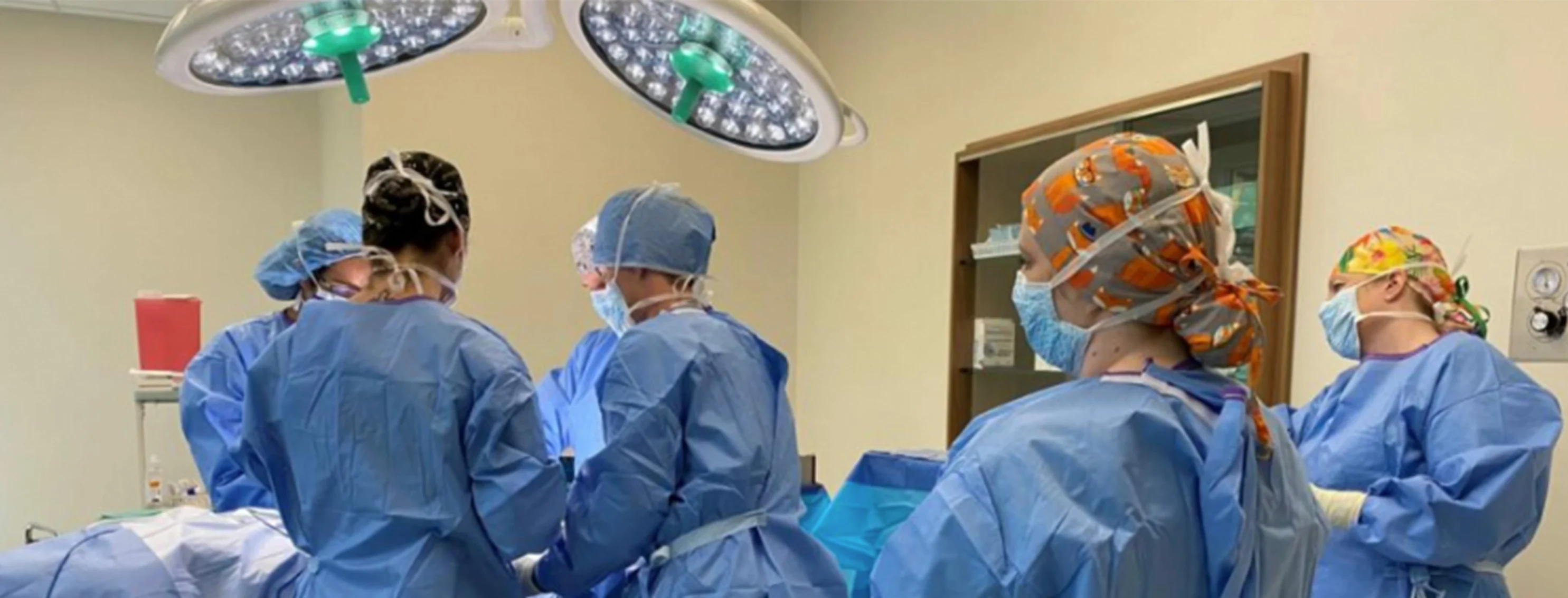
Medical therapy may be recommended in some patients; however, surgery is typically needed in most cases. Surgical removal of the gallbladder is the treatment of choice. In surgery the common bile duct is flushed of all mucinous bile to re-establish patency. In some cases, a stent (rubber tube) may be placed in the common bile duct to ensure continued flow of bile. At time of surgery the bile is cultured and a liver biopsy is taken to diagnose coinciding liver disease.

Perioperative mortality of gallbladder mucoceles ranges from 22% to 32%. A ruptured gallbladder has been shown to worsen the prognosis particularly when infection is present. Long-term survival of patients that have undergone gallbladder removal is excellent. Liver enzymes remain elevated in most patients, but these values usually are much lower after the healing process is completed. If the bile was infected patients typically need to stay on antibiotics for 4-6 weeks.

About the author:

Stephanie Szabo, DVM

Stephanie Szabo, DVM
Surgery Specialist at VETMED

Stephanie D. Szabo, VMD, DACVS obtained her veterinary degree from the University of Pennsylvania in 2003. She completed a general medicine and surgical internship at the Animal Medical Center in New York in 2004 and then went on to complete a second surgical internship at the Dallas Veterinary Surgical Center in 2005. After her internships she returned to the University of Pennsylvania in 2006 to complete a post-doctoral research fellowship in hip dysplasia. She then continued her training by completing a surgical residency at North Carolina State University in 2007.

After completing her residency she returned to Arizona in 2009 and became a Diplomate of the American College of Veterinary Surgeons in 2011.Dr. Stephanie Szabo is trained in all aspects of soft tissue, orthopedic, and neurologic surgery. She is also PennHIP certified and TTA (Tibial Tuberosity Advancement) trained. Her interests include emergency and trauma surgery, wound healing, and physical rehabilitation. She has been published in Veterinary Surgery, the Journal of the American Veterinary Medical Association, and the Journal of the American Animal Hospital Association.